Nasogastric feeding tubes play a crucial role in delivering enteral nutrition and medication to critically ill patients, yet their placement poses inherent risks and potential complications.
Previously, Levin tubes made of polyvinyl chloride were used primarily for drainage but were associated with complications such as gastrointestinal ulcers, bleeding, or perforation during prolonged use. Recent advancements in materials have led to the development of more flexible and softer silastic and polyurethane tubes, reducing the likelihood of gastrointestinal damage and enhancing patient comfort [
1,
2].
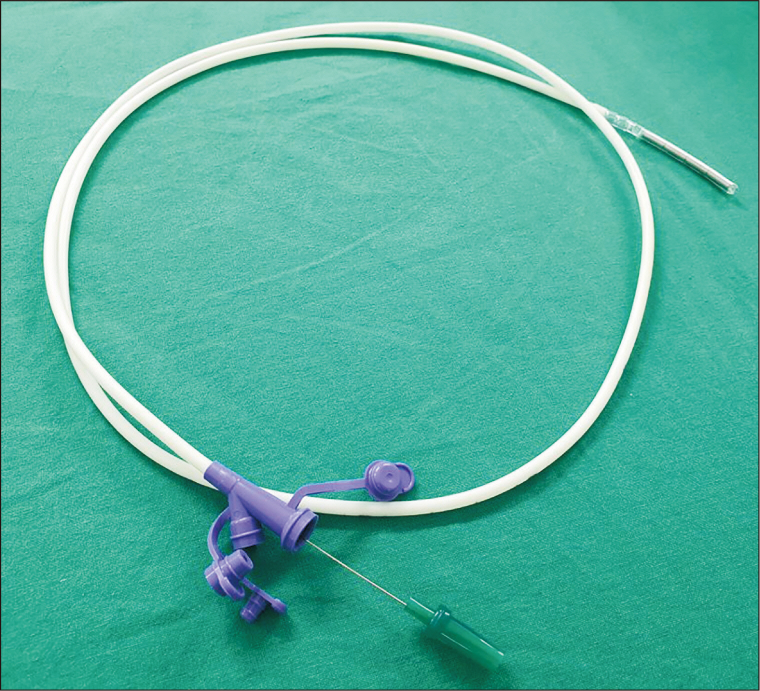
However, small-bore feeding tubes (
Fig. 1), which typically use a rigid guide wire during insertion, present significant risks, particularly in sedated, endotracheally intubated critically ill patients. In these cases, suppressed airway protection reflexes may lead to inadvertent airway insertion, resulting in various respiratory complications [
3,
4].
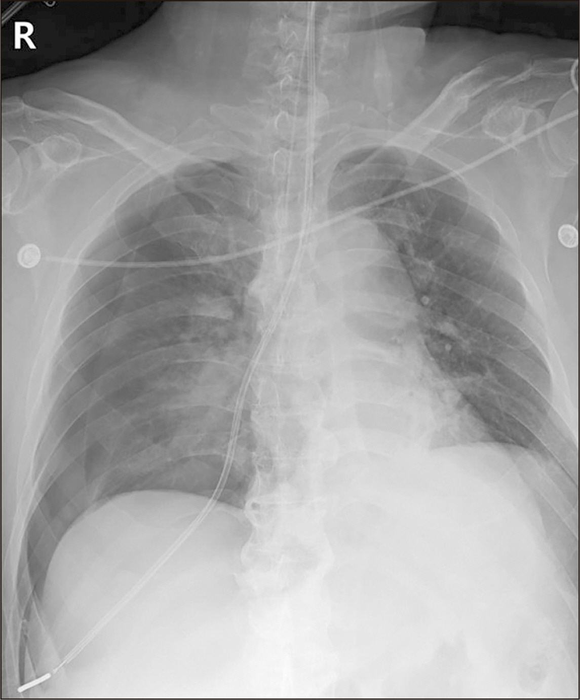
There have been several instances of pneumothorax due to improper positioning of small-bore nasogastric feeding tubes. A 60-year-old male patient in critical care following multiple injuries sustained from a traffic accident developed hypoxia immediately after nasogastric tube insertion, which was confirmed on chest X-ray to have punctured the lung parenchyma and entered the pleural space (
Fig. 2). The ensuing air leak required chest tube management for 7 days before resolution without further complications.
Therefore, during feeding tube insertion, operators must remain vigilant about the risk of malpositioning. Post-insertion confirmation of bubbling sounds through auscultation in three critical areas—specifically the left upper quadrant of the abdomen and both lungs—is crucial to ensure proper tube placement. This verification helps mitigate risks of malpositioning. The difficulty of localizing auscultatory sounds highlights the necessity of chest X-ray confirmation to precisely ascertain the tubés final position.
Multiple insertion techniques that could reduce the risk of malpositioning enteral feeding tubes have been investigated. These techniques include monitoring endotracheal cuff pressure to confirm proper tube insertion based on pressure changes, two-stage X-ray verification to ensure the tube tip is in the esophagus rather than the airway before advancing it into the stomach, post-insertion capnography measurement, real-time confirmation of tube position using fluoroscopy or endoscopy, and more recently, electromagnetic device-guided insertion techniques [
1]. However, these strategies may prolong insertion times, require additional equipment and specialized skills, and may not be efficient for critically ill patients in intensive care settings. Additionally, they may result in increased radiation exposure and higher costs, which are challenges to implementation in clinical practice.
In conclusion, while enteral nutrition via feeding tubes is crucial in critical care, caution is necessary to prevent unintended placement in the tracheobronchial tree, particularly in patients with endotracheal intubation or tracheotomy. Accurate determination of the feeding tube᾽s position post-insertion is imperative to minimize complications and optimize patient care.
Supplementary materials
None.
Acknowledgments
None.
Ethics statement
The informed consent was obtained from the patient.
Authors’ contribution
Conceptualization: SKH, HK. Resources: HK. Supervision: SKH. Writing – original draft: HK, Writing – review & editing: HK, SKH.
Conflict of interest
Suk-Kyung Hong is the Editor-in-Chief of the journal, but was not involved in the review process of this manuscript. Otherwise, there is no conflict of interest to disclose.
Funding
None.
Data availability
None.
Fig. 1Small-bore nasogastric feeding tube (EntriflexTM Nasogastric Feeding Tube; Covidien LLC).

Fig. 2Pneumothorax caused by malposition of an enteral feeding tube inserted in the right pleural space.

References
- 1. Choi KJ, Ryu JA, Park CM. Respiratory complications of small-bore feeding tube insertion in critically ill patients. J Acute Care Surg 2015;5:28-34. Article
- 2. Wendell GD, Lenchner GS, Promisloff RA. Pneumothorax complicating small-bore feeding tube placement. Arch Intern Med 1991;151:599-602. ArticlePubMed
- 3. Kolbitsch C, Pomaroli A, Lorenz I, Gassner M, Luger TJ. Pneumothorax following nasogastric feeding tube insertion in a tracheostomized patient after bilateral lung transplantation. Intensive Care Med 1997;23:440-2. ArticlePubMedPDF
- 4. Rassias AJ, Ball PA, Corwin HL. A prospective study of tracheopulmonary complications associated with the placement of narrow-bore enteral feeding tubes. Crit Care 1998;2:25-8. ArticlePubMedPMCPDF
Citations
Citations to this article as recorded by


 , Suk-Kyung Hong


 E-submission
 KSPEN
KSPEN KSSMN
KSSMN ASSMN
ASSMN JSSMN
JSSMN
 Cite
Cite

