Scopus, KCI, KoreaMed

Articles
- Page Path
- HOME > Ann Clin Nutr Metab > Volume 15(3); 2023 > Article
- Original Article Comparative assessment of nutritional characteristics of critically ill patients at admission and discharge from the neurosurgical intensive care unit in Korea: a comparison study
-
Eunjoo Bae1
 , Jinyoung Jang2
 , Miyeon Kim3
 , Seongsuk Kang3
 , Kumhee Son4,5
 , Taegon Kim2
 , Hyunjung Lim4,5
-
Annals of Clinical Nutrition and Metabolism 2023;15(3):97-108.
DOI: https://doi.org/10.15747/ACNM.2023.15.3.97
Published online: December 1, 2023
1Department of Food and Nutrition, CHA Bundang Medical Center, CHA University School of Medicine, Seongnam, Korea
2Department of Neurosurgery, CHA Bundang Medical Center, CHA University School of Medicine, Seongnam, Korea
3Department of Nursing, CHA Bundang Medical Center, CHA University School of Medicine, Seongnam, Korea
4Department of Medical Nutrition, Graduate School of East-West Medical Science, Kyung Hee University, Yongin, Korea
5Research Institute of Medical Nutrition, Kyung Hee University, Seoul, Korea
- Corresponding author: Hyunjung Lim, email: hjlim@khu.ac.kr
- Co-Corresponding author: Taegon Kim, email: tgkim@chamc.co.kr
© 2023 The Korean Society of Surgical Metabolism and Nutrition · The Korean Society for Parenteral and Enteral Nutrition
This is an open-access article distributed under the terms of the Creative Commons Attribution Non-Commercial License (http://creativecommons.org/licenses/by-nc/4.0), which permits unrestricted non-commercial use, distribution, and reproduction in any medium, provided the original work is properly cited.
- 3,284 Views
- 28 Download
- 2 Crossref
Abstract
-
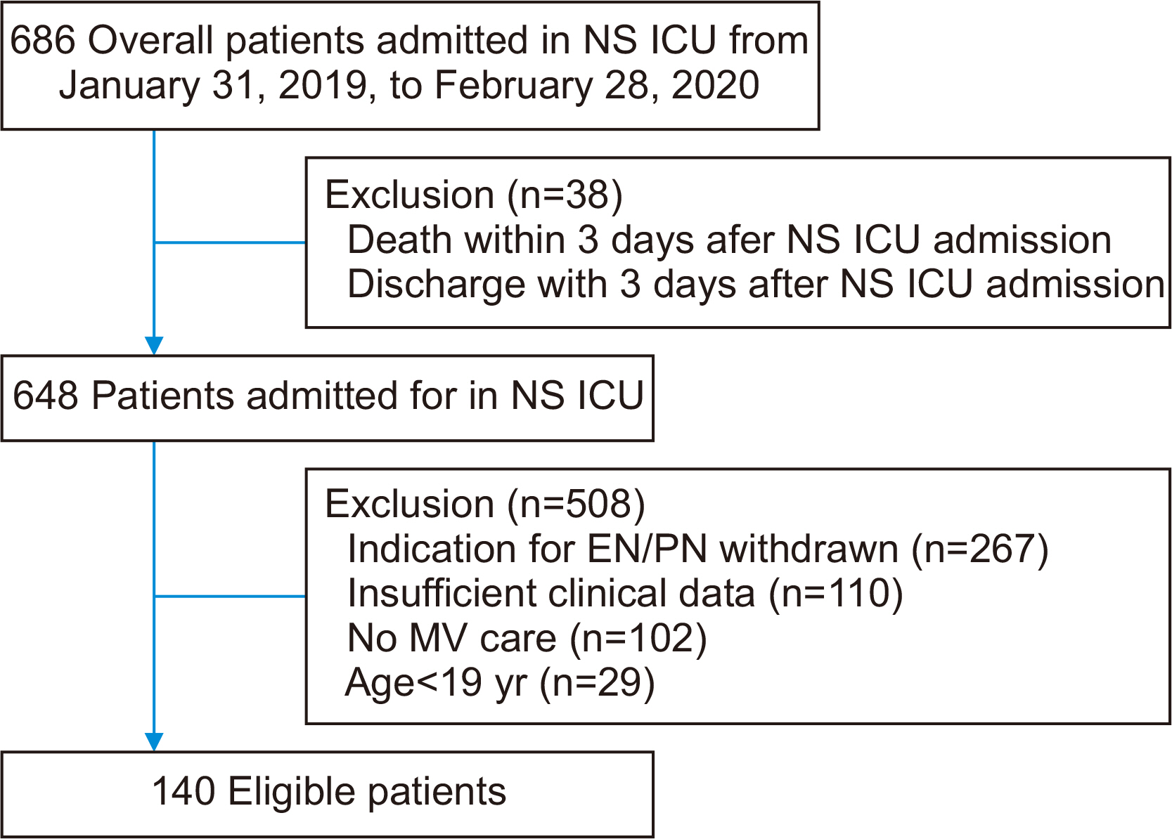
Purpose Patients in neurosurgical (NS) intensive care units (ICUs) experience considerable energy and protein deficits associated with adverse outcomes. This study aimed to compare the nutritional status of patients at admission to (baseline) and discharge from the NS ICU.
-
Methods This was a single-center, retrospective, before and after study of patients admitted in the NS ICU of the CHA Bundang Medical Center, from January 31, 2019, to February 28, 2020. All anthropometric data, biochemical data, clinical data, and dietary data were collected during the NS ICU stay. Specifically, we investigated the cumulative caloric deficit rate, phase angle and skeletal muscle index as indicators of lean muscle mass, and nitrogen balance according to demographic and clinical characteristics.
-
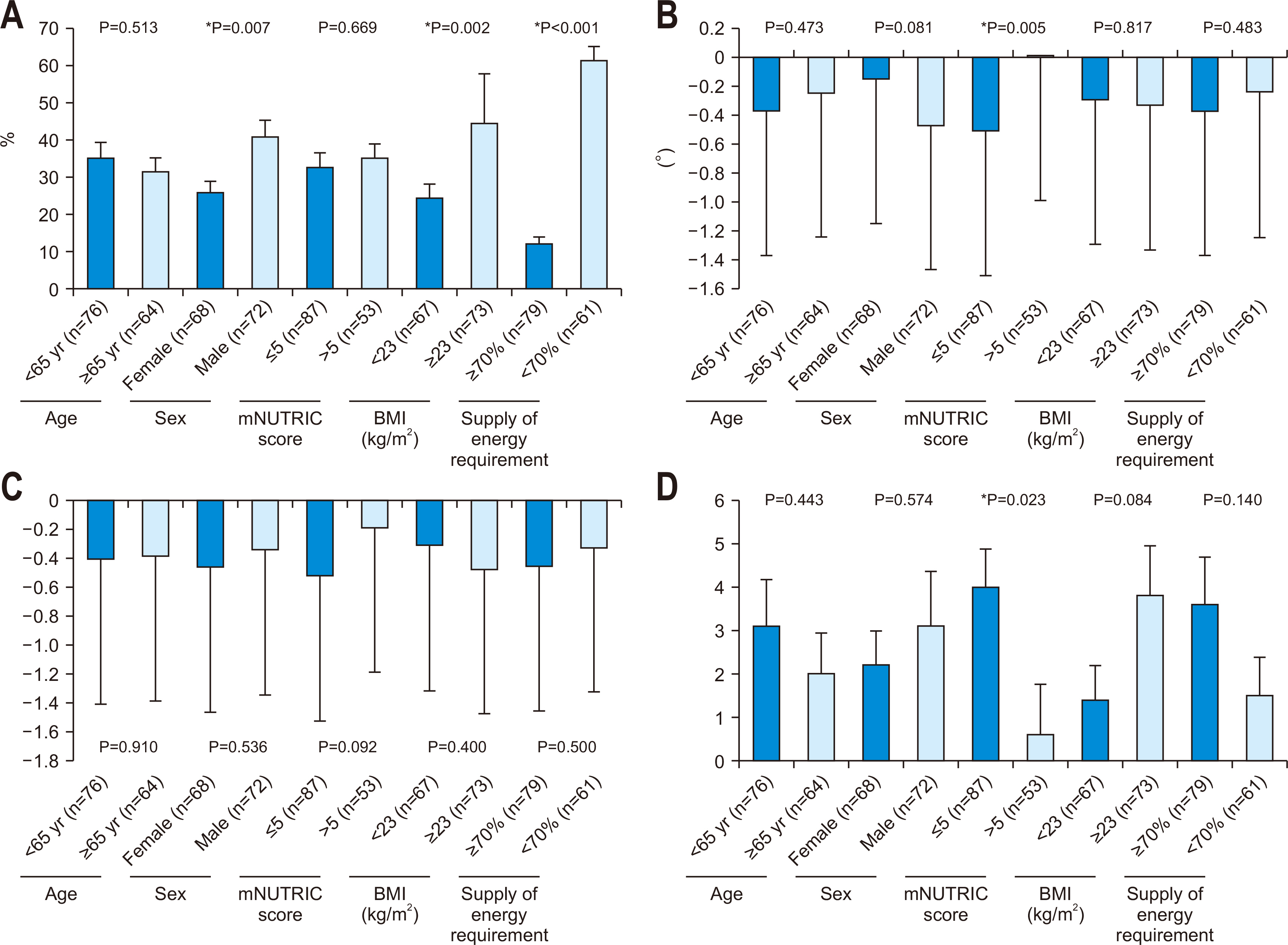
Results A total of 140 NS patients were studied. Calf circumference decreased from 31.4±4.2 cm at baseline to 30.2±4.0 cm at discharge (P<0.001). Energy supply rate increased from 44.4% at baseline to 89.2% at discharge. Phase angle (PhA) patients with an modified Nutrition Risk in the Critically ill (mNUTRIC) score≤5 group had significantly lower PhA values than those with an mNUTRIC score>5 (P=0.005).
-
Conclusion Although clinical and dietary parameters of patients in the NS ICU improved from baseline to discharge, anthropometric and biochemical markers of lean muscle mass and nutritional status decreased. PhA and nitrogen balance difference values were significantly different between those with an mNUTRIC score≤5 and those with an mNUTRIC score>5. These data indicate that the nutritional risk of critically ill patients increases during hospitalization in the NS ICU.
Introduction
Methods
Results
Discussion
Supplementary materials
Acknowledgments
Authors’ contribution
Conceptualization: EB, HL. Data curation: TK, JJ. Investigation: MK, SK. Methodology: EB, KS. Project administration: HL, TK. Resources: JJ, SK. Software: MK, KS. Supervision: HL, TK. Validation: JJ, SK. Visualization: EB, KS. Writing – original draft: EB, TK. Writing – review & editing: all authors.
Conflict of interest
The authors of this manuscript have no conflicts of interest to disclose.
Funding
None.
Data availability
Contact the corresponding author for data availability.


| Variable | All patients (n=140) |
|---|---|
| Age (yr) | 61.9±15.3 |
| Sex, male | 72 (51.4) |
| Height (cm) | 162.9±0.8 |
| mNUTRIC score | 5.1±1.4 |
| SAPS III score | 33.6±13.0 |
| Mechanical ventilator (day) | 9.5 (4.0–18.0) |
| Mechanical ventilator mode | |
| PCV | 80 (57.1) |
| PSIMV | 42 (30.0) |
| PSV | 13 (9.3) |
| CMV | 4 (2.9) |
| SIMV | 1 (0.7) |
| Primary diagnosis | |
| Brain hemorrhage | 101 (72.1) |
| Brain tumor | 25 (17.9) |
| Othera | 14 (10.0) |
| NS ICU LOS (day) | 21.0 (11.2–33.7) |
| Hospital LOS (day) | 39.5 (25.0–63.0) |
| NS ICU-free (day) | 14.0 (0.0–33.5) |
| Reason for NS ICU discharge | |
| Inter-hospital transfer | 90 (64.3) |
| Permanent care facility | 23 (16.4) |
| Death | 27 (19.3) |
| Surgeryb | 129 (92.1) |
| Comorbiditiesc | 106 (75.7) |
Values are presented as mean±standard deviation, number (%), or median (25th–75th percentiles).
mNUTRIC = modified Nutrition Risk in the Critically ill; SAPS = Simplified Acute Physiology Score; PCV = pressure-targeted controlled mandatory ventilation; PSIMV = pressure-targeted synchronized intermittent mandatory ventilation; PSV = pressure support ventilation; CMV = controlled mandatory ventilation; SIMV = synchronized intermittent mandatory ventilation; NS ICU = neurosurgical intensive care unit; LOS = length of stay.
aCerebral infarction, hydrocephalus meningitis, hypoxic brain damage, infective spondylopathies.
bExtraventricular drainage, craniotomy, burr hole, Guglielmi detachable coils embolization, intracerebral hemorrhage/catheter insert, craniectomy, decompression, ventriculo-other shunt, cerebral aneurysm clipping, operation of arteriovenous malformation, closed thoracostomy, tracheostomy.
cHypertension, diabetes mellitus, chronic renal failure, cancer, and others.
| Variable | Baseline (3rd day) | Discharge (last day) | P-value |
|---|---|---|---|
| Weight (kg) | 62.3±13.3 | 60.9±12.8 | <0.001* |
| Calf circumference (cm) | 31.4±4.2 | 30.2±4.0 | <0.001* |
| TSF (mm) | 19.3±7.4 | 19.5±7.2 | 0.276 |
| MAMC (cm) | 21.8±3.2 | 20.6±4.0 | <0.001* |
| MAMA (cm2) | 38.8±11.3 | 35.2±11.5 | <0.001* |
| BIA | |||
| Phase angle (°) | 4.0±1.4 | 3.7±1.3 | <0.001* |
| SMI | 5.7±1.1 | 5.3±1.1 | <0.001* |
| ECW/TBW | 0.401±0.015 | 0.405±0.014 | <0.001* |
| TBW/FFM | 74.0±0.54 | 74.0±0.57 | 0.892 |
| Serum albumin (g/dL) | 3.7±0.6 | 3.2±0.5 | <0.001* |
| Nitrogen balance | –8.5±5.2 | –5.8±5.7 | <0.001* |
| UUN (g/day) | 12.0±13.4 | 12.2±18.9 | 0.870 |
| APACHE II score | 22.6±5.1 | 20.8±6.6 | 0.001* |
| SOFA score | 5.8±1.9 | 5.1±3.1 | 0.005* |
| Glasgow Coma Scale | 5.2±3.0 | 6.4±3.9 | 0.001* |
| Braden Scale score | 13.0±3.1 | 13.0±2.6 | 0.816 |
| Pressure ulcer | 6 (4.3) | 33 (23.6) | 0.119 |
| Nutritional assessment | <0.001* | ||
| Adequately nourished status | 96 (68.6) | 63 (45.0) | |
| Mild malnutrition | 40 (28.6) | 69 (49.3) | |
| Moderate malnutrition | 2 (1.4) | 6 (4.3) | |
| Severe malnutrition | 2 (1.4) | 2 (1.4) |
Values are presented as mean±standard deviation or number (%).
TSF = triceps skinfold thickness; MAMC = mid-upper arm muscle circumference; MAMA = mid-upper arm muscle area; BIA = bioelectrical impedance analysis; SMI = skeletal muscle index; ECW = extracellular water; TBW = total body water; FFM = fat-free mass; UUN = 24-hour urine urea nitrogen; APACHE = Acute Physiology and Chronic Health Evaluation; SOFA = Sequential Organ Failure Assessment.
*Values are significantly different between intensive care unit baseline and discharge. P<0.05.
- 1. Annette H, Wenström Y. Implementing clinical guidelines for nutrition in a neurosurgical intensive care unit. Nurs Health Sci 2005;7:266-72. ArticlePubMed
- 2. Chapple LA, Chapman MJ, Lange K, Deane AM, Heyland DK. Nutrition support practices in critically ill head-injured patients: a global perspective. Crit Care 2016;20:6.ArticlePubMedPMCPDF
- 3. Mackay LE, Morgan AS, Bernstein BA. Factors affecting oral feeding with severe traumatic brain injury. J Head Trauma Rehabil 1999;14:435-47. ArticlePubMed
- 4. Chapple LS, Deane AM, Heyland DK, Lange K, Kranz AJ, Williams LT, et al. Energy and protein deficits throughout hospitalization in patients admitted with a traumatic brain injury. Clin Nutr 2016;35:1315-22. ArticlePubMed
- 5. Dhandapani SS, Manju D, Sharma BS, Mahapatra AK. Clinical malnutrition in severe traumatic brain injury: factors associated and outcome at 6 months. Indian J Neurotrauma 2007;4:35-9. Article
- 6. Wei X, Day AG, Ouellette-Kuntz H, Heyland DK. The association between nutritional adequacy and long-term outcomes in critically ill patients requiring prolonged mechanical ventilation: a multicenter cohort study. Crit Care Med 2015;43:1569-79. PubMed
- 7. Kress JP, Hall JB. ICU-acquired weakness and recovery from critical illness. N Engl J Med 2014;370:1626-35. ArticlePubMed
- 8. Abdelmalik PA, Dempsey S, Ziai W. Nutritional and bioenergetic considerations in critically ill patients with acute neurological injury. Neurocrit Care 2017;27:276-86. ArticlePubMedPDF
- 9. Costello LA, Lithander FE, Gruen RL, Williams LT. Nutrition therapy in the optimisation of health outcomes in adult patients with moderate to severe traumatic brain injury: findings from a scoping review. Injury 2014;45:1834-41. ArticlePubMed
- 10. Peterson SJ, Tsai AA, Scala CM, Sowa DC, Sheean PM, Braunschweig CL. Adequacy of oral intake in critically ill patients 1 week after extubation. J Am Diet Assoc 2010;110:427-33. ArticlePubMed
- 11. Pan WH, Yeh WT. How to define obesity? Evidence-based multiple action points for public awareness, screening, and treatment: an extension of Asian-Pacific recommendations. Asia Pac J Clin Nutr 2008;17:370-4. PubMed
- 12. de Vries MC, Koekkoek WK, Opdam MH, van Blokland D, van Zanten AR. Nutritional assessment of critically ill patients: validation of the modified NUTRIC score. Eur J Clin Nutr 2018;72:428-35. ArticlePubMedPDF
- 13. de Souza MFC, Zanei SSV, Whitaker IY. Risk of pressure injury in the ICU: transcultural adaptation and reliability of EVARUCI. Acta Paul Enferm 2018;31:201-8. Article
- 14. Swails WS, Samour PQ, Babineau TJ, Bistrian BR. A proposed revision of current ICD-9-CM malnutrition code definitions. J Am Diet Assoc 1996;96:370-3. ArticlePubMed
- 15. Alberda C, Gramlich L, Jones N, Jeejeebhoy K, Day AG, Dhaliwal R, et al. The relationship between nutritional intake and clinical outcomes in critically ill patients: results of an international multicenter observational study. Intensive Care Med 2009;35:1728-37. ArticlePubMedPDF
- 16. Tsai JR, Chang WT, Sheu CC, Wu YJ, Sheu YH, Liu PL, et al. Inadequate energy delivery during early critical illness correlates with increased risk of mortality in patients who survive at least seven days: a retrospective study. Clin Nutr 2011;30:209-14. ArticlePubMed
- 17. Peter JV, Moran JL, Phillips-Hughes J. A metaanalysis of treatment outcomes of early enteral versus early parenteral nutrition in hospitalized patients. Crit Care Med 2005;33:213-20. discussion 260-1. ArticlePubMed
- 18. Hafsteinsdóttir TB, Mosselman M, Schoneveld C, Riedstra YD, Kruitwagen CL. Malnutrition in hospitalised neurological patients approximately doubles in 10 days of hospitalisation. J Clin Nurs 2010;19:639-48. ArticlePubMed
- 19. Deutschman CS, Konstantinides FN, Raup S, Thienprasit P, Cerra FB. Physiological and metabolic response to isolated closed-head injury. Part 1: basal metabolic state: correlations of metabolic and physiological parameters with fasting and stressed controls. J Neurosurg 1986;64:89-98. PubMed
- 20. Hoffer LJ. Protein and energy provision in critical illness. Am J Clin Nutr 2003;78:906-11. ArticlePubMed
- 21. Kreymann G, DeLegge MH, Luft G, Hise ME, Zaloga GP. The ratio of energy expenditure to nitrogen loss in diverse patient groups--a systematic review. Clin Nutr 2012;31:168-75. ArticlePubMed
- 22. Wirth R, Volkert D, Rösler A, Sieber CC, Bauer JM. Bioelectric impedance phase angle is associated with hospital mortality of geriatric patients. Arch Gerontol Geriatr 2010;51:290-4. ArticlePubMed
- 23. Thibault R, Makhlouf AM, Mulliez A, Cristina Gonzalez M, Kekstas G, Kozjek NR, et al. Phase Angle Project Investigators. Fat-free mass at admission predicts 28-day mortality in intensive care unit patients: the international prospective observational study Phase Angle Project. Intensive Care Med 2016;42:1445-53. ArticlePubMedPDF
- 24. Berger MM, Reintam-Blaser A, Calder PC, Casaer M, Hiesmayr MJ, Mayer K, et al. Monitoring nutrition in the ICU. Clin Nutr 2019;38:584-93. ArticlePubMed
- 25. Kondrup J, Allison SP, Elia M, Vellas B, Plauth M. ESPEN guidelines for nutrition screening 2002. Clin Nutr 2003;22:415-21. ArticlePubMed
- 26. Kondrup J, Rasmussen HH, Hamberg O, Stanga Z. Ad Hoc ESPEN Working Group. Nutritional risk screening (NRS 2002): a new method based on an analysis of controlled clinical trials. Clin Nutr 2003;22:321-36. ArticlePubMed
- 27. Simpson F, Doig GS. Early PN Trial Investigators Group. Physical assessment and anthropometric measures for use in clinical research conducted in critically ill patient populations: an analytic observational study. JPEN J Parenter Enteral Nutr 2015;39:313-21. PubMed
- 28. Sheean PM, Peterson SJ, Gurka DP, Braunschweig CA. Nutrition assessment: the reproducibility of subjective global assessment in patients requiring mechanical ventilation. Eur J Clin Nutr 2010;64:1358-64. ArticlePubMedPMCPDF
- 29. Miyajima I, Yatabe T, Kuroiwa H, Tamura T, Yokoyama M. Influence of nutrition support therapy on readmission among patients with acute heart failure in the intensive care unit: a single-center observational study. Clin Nutr 2020;39:174-9. ArticlePubMed
- 30. Sungurtekin H, Sungurtekin U, Oner O, Okke D. Nutrition assessment in critically ill patients. Nutr Clin Pract 2008;23:635-41. ArticlePubMedPDF
- 31. Kim D, Sun JS, Lee YH, Lee JH, Hong J, Lee JM. Comparative assessment of skeletal muscle mass using computerized tomography and bioelectrical impedance analysis in critically ill patients. Clin Nutr 2019;38:2747-55. ArticlePubMed
- 32. Rogenski NM, Santos VL. [Incidence of pressure ulcers at a university hospital]. Rev Lat Am Enfermagem 2005;13:474-80; Portuguese. PubMed
- 33. Blanes L, Duarte IS, Calil JA, Ferriera LM. Clinical and epidemiological assessment of pressure ulcers in patients admitted to Sao Paulo Hospital. Rev Assoc Méd Bras 2004;50:182-7.
- 34. Hyun S, Li X, Vermillion B, Newton C, Fall M, Kaewprag P, et al. Body mass index and pressure ulcers: improved predictability of pressure ulcers in intensive care patients. Am J Crit Care 2014;23:494-500. quiz 501.ArticlePubMedPMC
- 35. Becker D, Tozo TC, Batista SS, Mattos AL, Silva MCB, Rigon S, et al. Pressure ulcers in ICU patients: incidence and clinical and epidemiological features: a multicenter study in southern Brazil. Intensive Crit Care Nurs 2017;42:55-61. ArticlePubMed
- 36. Kaya H, Turan N, Özdemir Aydın G. Stan odżywienia pacjentów neurochirurgicznego oddziału intensywnej opieki medycznej. J Neurol Neurosurg Nurs 2017;6:33-8; Polish. ArticlePDF
- 37. Heyland DK, Dhaliwal R, Jiang X, Day AG. Identifying critically ill patients who benefit the most from nutrition therapy: the development and initial validation of a novel risk assessment tool. Crit Care 2011;15:R268.ArticlePubMedPMCPDF
- 38. Mukhopadhyay A, Henry J, Ong V, Leong CS, Teh AL, van Dam RM, et al. Association of modified NUTRIC score with 28-day mortality in critically ill patients. Clin Nutr 2017;36:1143-8. ArticlePubMed
- 39. Ardehali SH, Dehghan S, Baghestani AR, Velayati A, Vahdat Shariatpanahi Z. Association of admission serum levels of vitamin D, calcium, Phosphate, magnesium and parathormone with clinical outcomes in neurosurgical ICU patients. Sci Rep 2018;8:2965.ArticlePubMedPMCPDF
- 40. Dionyssiotis Y, Papachristos A, Petropoulou K, Papathanasiou J, Papagelopoulos P. Nutritional alterations associated with neurological and neurosurgical diseases. Open Neurol J 2016;10:32-41. ArticlePubMedPMCPDF
References
Figure & Data
REFERENCES
Citations

- A Review on the Effects of Multiple Nutritional Scores on Wound Healing after Neurosurgery.
Jingqian Ye, Bo Ning , Jianwen Zhi
International Journal of Biology and Life Sciences.2025; 9(2): 82. CrossRef - Transition from Enteral to Oral Nutrition in Intensive Care and Post Intensive Care Patients: A Scoping Review
Gioia Vinci, Nataliia Yakovenko, Elisabeth De Waele, Reto Stocker
Nutrients.2025; 17(11): 1780. CrossRef


Fig. 1
Fig. 2
Baseline demographic and clinical characteristics
| Variable | All patients (n=140) |
|---|---|
| Age (yr) | 61.9±15.3 |
| Sex, male | 72 (51.4) |
| Height (cm) | 162.9±0.8 |
| mNUTRIC score | 5.1±1.4 |
| SAPS III score | 33.6±13.0 |
| Mechanical ventilator (day) | 9.5 (4.0–18.0) |
| Mechanical ventilator mode | |
| PCV | 80 (57.1) |
| PSIMV | 42 (30.0) |
| PSV | 13 (9.3) |
| CMV | 4 (2.9) |
| SIMV | 1 (0.7) |
| Primary diagnosis | |
| Brain hemorrhage | 101 (72.1) |
| Brain tumor | 25 (17.9) |
| Other |
14 (10.0) |
| NS ICU LOS (day) | 21.0 (11.2–33.7) |
| Hospital LOS (day) | 39.5 (25.0–63.0) |
| NS ICU-free (day) | 14.0 (0.0–33.5) |
| Reason for NS ICU discharge | |
| Inter-hospital transfer | 90 (64.3) |
| Permanent care facility | 23 (16.4) |
| Death | 27 (19.3) |
| Surgery |
129 (92.1) |
| Comorbidities |
106 (75.7) |
Values are presented as mean±standard deviation, number (%), or median (25th–75th percentiles).
mNUTRIC = modified Nutrition Risk in the Critically ill; SAPS = Simplified Acute Physiology Score; PCV = pressure-targeted controlled mandatory ventilation; PSIMV = pressure-targeted synchronized intermittent mandatory ventilation; PSV = pressure support ventilation; CMV = controlled mandatory ventilation; SIMV = synchronized intermittent mandatory ventilation; NS ICU = neurosurgical intensive care unit; LOS = length of stay.
aCerebral infarction, hydrocephalus meningitis, hypoxic brain damage, infective spondylopathies.
bExtraventricular drainage, craniotomy, burr hole, Guglielmi detachable coils embolization, intracerebral hemorrhage/catheter insert, craniectomy, decompression, ventriculo-other shunt, cerebral aneurysm clipping, operation of arteriovenous malformation, closed thoracostomy, tracheostomy.
cHypertension, diabetes mellitus, chronic renal failure, cancer, and others.
Anthropometric, biochemical, and clinical data at intensive care unit baseline and discharge
| Variable | Baseline (3rd day) | Discharge (last day) | P-value |
|---|---|---|---|
| Weight (kg) | 62.3±13.3 | 60.9±12.8 | <0.001 |
| Calf circumference (cm) | 31.4±4.2 | 30.2±4.0 | <0.001 |
| TSF (mm) | 19.3±7.4 | 19.5±7.2 | 0.276 |
| MAMC (cm) | 21.8±3.2 | 20.6±4.0 | <0.001 |
| MAMA (cm2) | 38.8±11.3 | 35.2±11.5 | <0.001 |
| BIA | |||
| Phase angle (°) | 4.0±1.4 | 3.7±1.3 | <0.001 |
| SMI | 5.7±1.1 | 5.3±1.1 | <0.001 |
| ECW/TBW | 0.401±0.015 | 0.405±0.014 | <0.001 |
| TBW/FFM | 74.0±0.54 | 74.0±0.57 | 0.892 |
| Serum albumin (g/dL) | 3.7±0.6 | 3.2±0.5 | <0.001 |
| Nitrogen balance | –8.5±5.2 | –5.8±5.7 | <0.001 |
| UUN (g/day) | 12.0±13.4 | 12.2±18.9 | 0.870 |
| APACHE II score | 22.6±5.1 | 20.8±6.6 | 0.001 |
| SOFA score | 5.8±1.9 | 5.1±3.1 | 0.005 |
| Glasgow Coma Scale | 5.2±3.0 | 6.4±3.9 | 0.001 |
| Braden Scale score | 13.0±3.1 | 13.0±2.6 | 0.816 |
| Pressure ulcer | 6 (4.3) | 33 (23.6) | 0.119 |
| Nutritional assessment | <0.001 |
||
| Adequately nourished status | 96 (68.6) | 63 (45.0) | |
| Mild malnutrition | 40 (28.6) | 69 (49.3) | |
| Moderate malnutrition | 2 (1.4) | 6 (4.3) | |
| Severe malnutrition | 2 (1.4) | 2 (1.4) |
Values are presented as mean±standard deviation or number (%).
TSF = triceps skinfold thickness; MAMC = mid-upper arm muscle circumference; MAMA = mid-upper arm muscle area; BIA = bioelectrical impedance analysis; SMI = skeletal muscle index; ECW = extracellular water; TBW = total body water; FFM = fat-free mass; UUN = 24-hour urine urea nitrogen; APACHE = Acute Physiology and Chronic Health Evaluation; SOFA = Sequential Organ Failure Assessment.
*Values are significantly different between intensive care unit baseline and discharge. P<0.05.
Nutritional targets and deficits during neurosurgical intensive care unit hospitalization
| Variable | All patients (n=140) |
|---|---|
| Days from NS ICU admission to initiating nutrition support | 1.75±1.0 |
| Patients who started EN within 48 hours from NS ICU admission | 61 (43.6) |
| Days receiving EN | 20.2±17.2 |
| Targets | |
| Calories prescribed (kcal/day) | 1,600 (1,300–1,700) |
| Calories prescribed (kcal/kg) | 24.6 (24.1–25.7) |
| Total energy requirements during NS ICU period (kcal) | 31,500 (15,400–54,150) |
| Proteins prescribed (g/day) | 80 (70–90) |
| Proteins prescribed (g/kg) | 1.26 (1.23–1.30) |
| Total protein requirements during NS ICU period (g) | 1,617 (785–2,730) |
| Deficits | |
| Cumulative caloric deficit (kcal) | 8,571 (3,232–14,362) |
| % Cumulative caloric deficit | 27.9 (13.3–48.0) |
| Cumulative protein deficit (g) | 516.9 (210.3–840.6) |
| % Cumulative protein deficit | 33.2 (16.0–51.8) |
Values are presented as mean±standard deviation, number (%), or median (25th–75th percentiles).
NS ICU = neurosurgical intensive care unit; EN = enteral nutrition.
Nutrition provision comparisons between intensive care unit baseline and discharge
| Variable | Baseline (3rd day) | Discharge (last day) |
|---|---|---|
| Calorie intake | ||
| Calories received (kcal/day) | 659.0 (421–861) | 1,354.6 (1,119–1,546) |
| Calories received (kcal/kg) | 10.1 (7.1–14.1) | 23.5 (18.2–25.9) |
| % Calories received of prescribed | 42.0 (28.4–57.1) | 95.3 (74.0–106.6) |
| % Enteral nutrition of calories prescribed | 0 (0–37.5) | 92.8 (69.4–100) |
| % Parenteral nutrition of calories prescribed | 22.0 (9.5–35.5) | 0 (0–0.5) |
| Daily caloric deficit (kcal/day) | 840.3 (608.7–1,183.1) | 77.0 (–100.0 to 400.0) |
| Protein intake | ||
| Protein received (g/day) | 35.1 (23.5–46.5) | 60.9 (50.0–72.0) |
| Protein received (g/kg) | 0.5 (0.3–0.7) | 1.0 (0.8–1.2) |
| % Protein received of prescribed | 45.7 (29.4–59.6) | 83.0 (68.0–101.6) |
| % Enteral nutrition of protein prescribed | 0 (0–34.1) | 80.0 (60.0–96.2) |
| % Parenteral nutrition of protein prescribed | 29.4 (15.7–45.7) | 0 (0–0) |
| Daily protein deficit (g/day) | 42.7 (29.0–58.9) | 12.0 (–1.0 to 26.0) |
Values are presented as median (25th–75th percentiles).
P-values of all data by paired t-test were <0.001.
Multiple linear regression analysis of cumulative caloric deficit% during neurosurgical intensive care unit hospitalization (
| Independent variable | β (standardized) | SE | t-value | P-value |
|---|---|---|---|---|
| Constant | 50.736 | 5.035 | 10.078 | <0.001 |
| Age≥65 yr | 7.509 (0.111) | 4.260 | 1.763 | 0.080 |
| Sex, male | 6.004 (0.089) | 3.966 | 1.514 | 0.132 |
| mNUTRIC>5 | –6.748 (–0.097) | 4.256 | –1.585 | 0.115 |
| Body mass index≥23 kg/m2 | 11.961 (0.177) | 3.890 | 3.075 | 0.003 |
| Supply of energy requirement≥70% | –48.601 (–0.715) | 3.990 | –12.180 | <0.001 |
SE = standard error; mNUTRIC = modified Nutrition Risk in the Critically ill.
aThe suitability of the regression model for the 5 independent variables was confirmed through the P-value of the ANOVA result table.
Multiple linear regression analysis of phase angle difference between intensive care unit baseline and discharge (
| Independent variable | β (standardized) | SE | t-value | P-value |
|---|---|---|---|---|
| Constant | –0.210 | 0.236 | –0.890 | 0.375 |
| Age≥65 yr | –0.134 (–0.062) | 0.200 | –0.672 | 0.503 |
| Sex, male | –0.364 (–0.169) | 0.186 | –1.953 | 0.053 |
| mNUTRIC>5 | 0.552 (0.249) | 0.200 | 2.761 | 0.007 |
| Body mass index≥23 kg/m2 | –0.008 (–0.003) | 0.183 | –0.041 | 0.967 |
| Supply of energy requirement≥70% | –0.107 (–0.049) | 0.187 | –0.571 | 0.569 |
SE = standard error; mNUTRIC = modified Nutrition Risk in the Critically ill.
aThe suitability of the regression model for the 5 independent variables was confirmed through the P-value of the ANOVA result table.
Multiple linear regression analysis of skeletal muscle index difference between intensive care unit baseline and discharge (
| Independent variable | β (standardized) | SE | t-value | P-value |
|---|---|---|---|---|
| Constant | –0.407 | 0.259 | –1.574 | 0.118 |
| Age≥65 yr | –0.099 (–0.043) | 0.219 | –0.451 | 0.653 |
| Sex, male | 0.111 (0.049) | 0.204 | 0.546 | 0.586 |
| mNUTRIC>5 | 0.353 (0.150) | 0.219 | 1.615 | 0.109 |
| Body mass index≥23 kg/m2 | –0.189 (–0.082) | 0.200 | –0.945 | 0.347 |
| Supply of energy requirement≥70% | –0.073 (–0.032) | 0.205 | –0.356 | 0.723 |
SE = standard error; mNUTRIC = modified Nutrition Risk in the Critically ill.
aThe suitability of the regression model for the 5 independent variables was confirmed through the P-value of the ANOVA result table.
Multiple linear regression analysis of nitrogen balance difference between intensive care unit baseline and discharge (
| Independent variable | β (standardized) | SE | t-value | P-value |
|---|---|---|---|---|
| Constant | 2.276 | 1.518 | 1.500 | 0.136 |
| Age≥65 yr | 0.542 (0.040) | 1.284 | 0.422 | 0.674 |
| Sex, male | 0.644 (0.047) | 1.196 | 0.539 | 0.591 |
| mNUTRIC>5 | –2.552 (–0.183) | 1.283 | –1.989 | 0.049 |
| Body mass index≥23 kg/m2 | 1.689 (0.124) | 1.172 | 1.440 | 0.152 |
| Supply of energy requirement≥70% | –0.324 (–0.024) | 1.203 | –0.269 | 0.788 |
SE = standard error; mNUTRIC = modified Nutrition Risk in the Critically ill.
aThe suitability of the regression model for the 5 independent variables was confirmed through the P-value of the ANOVA result table.
Values are presented as mean±standard deviation, number (%), or median (25th–75th percentiles). mNUTRIC = modified Nutrition Risk in the Critically ill; SAPS = Simplified Acute Physiology Score; PCV = pressure-targeted controlled mandatory ventilation; PSIMV = pressure-targeted synchronized intermittent mandatory ventilation; PSV = pressure support ventilation; CMV = controlled mandatory ventilation; SIMV = synchronized intermittent mandatory ventilation; NS ICU = neurosurgical intensive care unit; LOS = length of stay. aCerebral infarction, hydrocephalus meningitis, hypoxic brain damage, infective spondylopathies. bExtraventricular drainage, craniotomy, burr hole, Guglielmi detachable coils embolization, intracerebral hemorrhage/catheter insert, craniectomy, decompression, ventriculo-other shunt, cerebral aneurysm clipping, operation of arteriovenous malformation, closed thoracostomy, tracheostomy. cHypertension, diabetes mellitus, chronic renal failure, cancer, and others.
Values are presented as mean±standard deviation or number (%). TSF = triceps skinfold thickness; MAMC = mid-upper arm muscle circumference; MAMA = mid-upper arm muscle area; BIA = bioelectrical impedance analysis; SMI = skeletal muscle index; ECW = extracellular water; TBW = total body water; FFM = fat-free mass; UUN = 24-hour urine urea nitrogen; APACHE = Acute Physiology and Chronic Health Evaluation; SOFA = Sequential Organ Failure Assessment. *Values are significantly different between intensive care unit baseline and discharge. P<0.05.
Values are presented as mean±standard deviation, number (%), or median (25th–75th percentiles). NS ICU = neurosurgical intensive care unit; EN = enteral nutrition.
Values are presented as median (25th–75th percentiles). P-values of all data by paired t-test were <0.001.
SE = standard error; mNUTRIC = modified Nutrition Risk in the Critically ill. aThe suitability of the regression model for the 5 independent variables was confirmed through the P-value of the ANOVA result table.
SE = standard error; mNUTRIC = modified Nutrition Risk in the Critically ill. aThe suitability of the regression model for the 5 independent variables was confirmed through the P-value of the ANOVA result table.
SE = standard error; mNUTRIC = modified Nutrition Risk in the Critically ill. aThe suitability of the regression model for the 5 independent variables was confirmed through the P-value of the ANOVA result table.
SE = standard error; mNUTRIC = modified Nutrition Risk in the Critically ill. aThe suitability of the regression model for the 5 independent variables was confirmed through the P-value of the ANOVA result table.

 E-submission
 KSPEN
KSPEN KSSMN
KSSMN ASSMN
ASSMN JSSMN
JSSMN Cite
Cite

